Identifies organelle regional localization and migration within the yeast cell 3D space in snapshot/timeseries data. Pipeline works in tandem with our cell segmentation and classification utility.
All cells that are pushed through this pipeline are standardized in origin and axes placement to allow for comparison of structure localization between cells. All organelles within a cell are aligned to a pre-defined axis system, which is defined as follows:
For Single (un-budded) Yeast Cells
Attributes measureable for un-budded yeast cells are as follows:
- Cross sectional area
- Volume
- Perimeter
- Major axis length
- Minor axis length
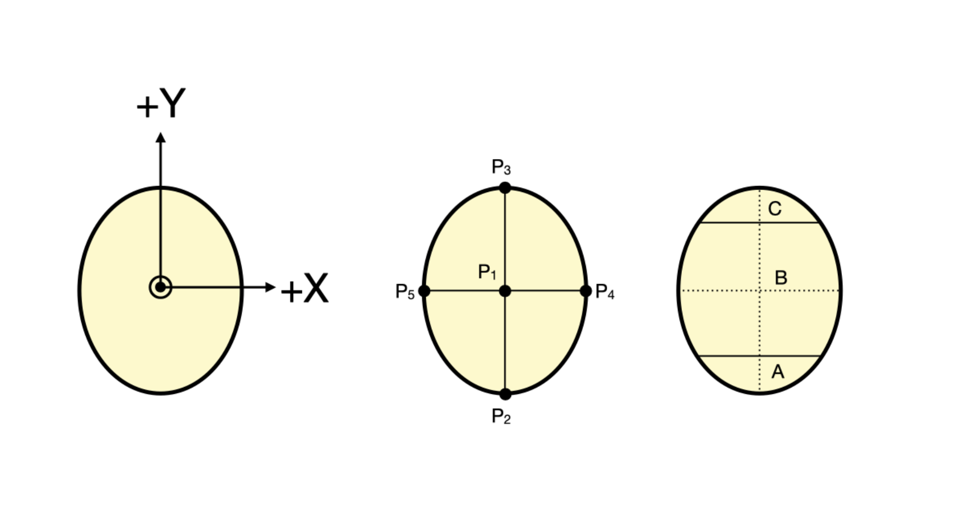
For organelle tracking, each unbudded cell is the shape of a prolate spheroid with the center of the cell is defined as the origin, the Y axis defined as parallel to the major axis length, and X axis defined as parallel to the minor axis. Each cell is presumed to be radially symmetric about its major axis. The Z axis is defined as in/out of the page.
Localization of organelles in regions A,B,C (see diagram above) can also be determined, with respect to the same origin (P1).
For Budded Yeast Cells
Attributes measureable for both the bud and mother portion of the budding cell:
- Cross sectional area
- Volume
- Perimeter
- Major axis length
- Minor axis length
Additionally, the following attributes are also included:
- Budding neck diameter (N1-N2)
- Direct Pole-to-pole distance from mother apex to bud apex (P1 to P5 directly)
- Indirect Pole-to-pole distance from mother apex to bud apex, through the center of the bud neck (P1-P2-P3-P4-P5)
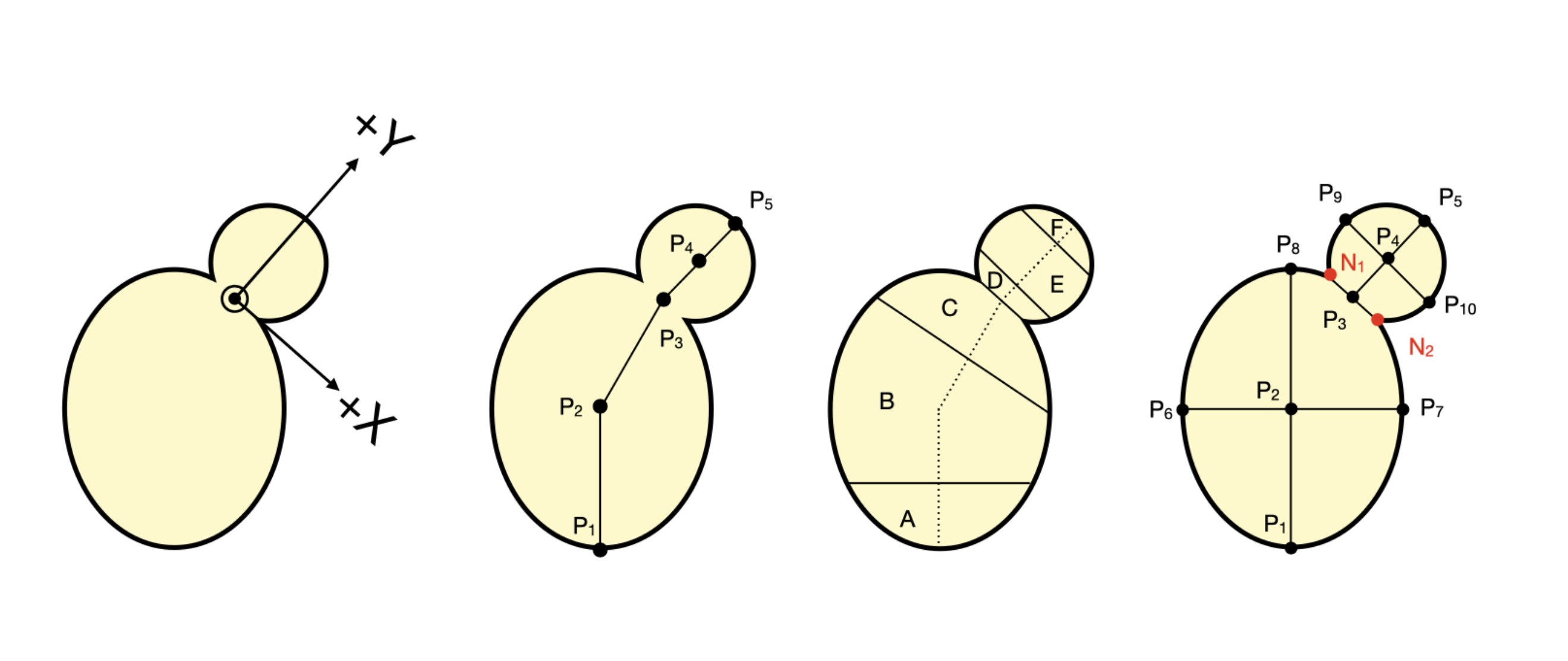
For organelle tracking, each budding cell is defined as two prolate spheroids fused together, with the smaller spheroid forming the bud and the larger the mother. The origin is defined as the center of the bud neck, with the X axis direction defined as parallel to the axis that forms the bud neck, and the +Y direction in the direction towards the bud and orthogonal to the X axis, intersecting at the bud neck center. The Z axis is defined as in/out of the page.
Localization of organelles in regions A,B,C,D,E,F (see diagram above) can also be determined, with respect to the same origin (P2).
Mapping of points on the cell structure is retained in output files as seen below (can be double checked in ImageJ):